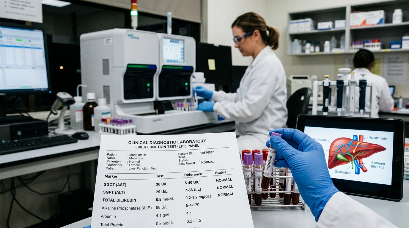
The liver is the largest internal solid organ in the human body, serving as an unmatched biochemical powerhouse responsible for over 500 essential physiological functions. It filters portal blood coming from the digestive tract, metabolizes carbohydrates and fats, neutralizes drugs and environmental toxins, synthesizes essential blood-clotting proteins and albumin, and produces bile to digest dietary lipids. Because liver tissue does not contain sensory nerve fibers within its functional parenchyma, liver inflammation, cellular swelling, and fat accumulation progress silently without pain. A Liver Function Test (LFT Panel) is the primary diagnostic tool used by clinicians to detect hepatocellular injury before permanent fibrotic remodeling occurs.
What Is a Liver Function Test?
A Liver Function Test is a panel of biochemical blood assays that measure two distinct aspects of hepatic biology:
- Hepatocellular Integrity Markers (Enzymes): Measures intracellular enzymes that leak into the bloodstream when liver cells (hepatocytes) suffer membrane damage or necrosis.
- Hepatic Functional Capacity (Proteins & Bilirubin): Measures the liver's synthetic ability to manufacture essential proteins (Albumin and Globulin) and its excretory capability to clear and conjugate Bilirubin via the biliary tree.
Hepatic Enzymes & Synthetic Biomarkers Explained
A comprehensive LFT panel evaluates several specific parameters:
1. SGPT (ALT - Alanine Aminotransferase)
ALT is located primarily inside the cytoplasm of liver hepatocytes. Because it exists in very low concentrations in other tissues, an elevation in SGPT is the most specific biomarker for acute or chronic liver inflammation (hepatitis, fatty liver, medication toxicity).
2. SGOT (AST - Aspartate Aminotransferase)
AST is found in liver cells, but is also present in cardiac muscle, skeletal muscle, kidneys, and brain tissue. While elevated AST reflects liver injury, it can also rise following intense muscle trauma or acute myocardial stress.
3. Alkaline Phosphatase (ALP) & Gamma-Glutamyl Transferase (GGT)
ALP is an enzyme concentrated in the epithelial lining of the bile ducts, as well as in bone and placenta. An elevated ALP with normal ALT/AST points toward biliary obstruction (gallstones, biliary strictures) or bone disorders. GGT is a sensitive marker of biliary epithelial stress and chronic alcohol exposure.
4. Bilirubin (Total, Direct, and Indirect)
Bilirubin is the yellow-orange pigment produced during the normal breakdown of aged red blood cells. The spleen produces Indirect (Unconjugated) Bilirubin, which travels to the liver, where hepatocytes conjugate it with glucuronic acid to form water-soluble Direct (Conjugated) Bilirubin for excretion in bile. High total bilirubin (> 2.0 mg/dL) causes visible jaundice (yellowing of sclera and skin).
5. Total Protein, Albumin, Globulin, and A/G Ratio
Albumin is manufactured exclusively by the liver and accounts for over 50% of total plasma protein, maintaining oncotic pressure to prevent fluid leaking into tissues. A low serum albumin (< 3.5 g/dL) is a hallmark of chronic, advanced liver impairment or severe malnutrition.
Master LFT Reference Ranges & Clinical Significance
The following table outlines standard clinical reference intervals and interpretations for the complete LFT panel:
| Parameter | Standard Reference Range | Clinical Meaning of Elevated Levels | Clinical Meaning of Depleted Levels |
|---|---|---|---|
| SGPT (ALT) | 7–56 U/L | Fatty liver (NAFLD), viral hepatitis (A, B, C, E), alcohol injury, drug-induced liver injury (DILI). | Normal baseline state. |
| SGOT (AST) | 10–40 U/L | Alcoholic hepatitis, viral hepatitis, cirrhosis, intense muscle exertion, myocardial infarction. | Normal baseline state. |
| Total Bilirubin | 0.2–1.2 mg/dL | Hemolytic anemia, Gilbert syndrome, viral hepatitis, gallstone obstruction, cirrhosis. | No clinical significance. |
| Direct Bilirubin | 0.0–0.3 mg/dL | Biliary obstruction (choledocholithiasis, biliary stricture), acute hepatitis, drug cholestasis. | Normal baseline state. |
| Alkaline Phosphatase | 44–147 U/L | Biliary obstruction, cholestasis, bone metastases, Paget disease, rapid adolescent bone growth. | Severe zinc deficiency, malnutrition. |
| Serum Albumin | 3.5–5.0 g/dL | Dehydration (hemoconcentration). | Chronic liver cirrhosis (impaired synthesis), nephrotic syndrome (renal leakage), severe protein malnutrition. |
| A/G Ratio | 1.2–2.2 | Normal physiological state. | Reversal (< 1.0) is a classic indicator of chronic active cirrhosis or multiple myeloma. |
Non-Alcoholic Fatty Liver Disease (NAFLD) & LFT Findings
Non-Alcoholic Fatty Liver Disease (NAFLD / MASLD) has become the most common chronic liver condition worldwide, affecting up to 35% of Indian adults. In NAFLD, excessive consumption of high-fructose corn syrup, refined carbohydrates, and saturated fats leads to ectopic lipid accumulation within hepatocytes.
On routine blood tests, NAFLD presents with mild-to-moderate elevations in SGPT (50–120 U/L) and SGOT, often accompanied by elevated Triglycerides and borderline fasting glucose. Confirming NAFLD with an abdominal ultrasound or transient elastography (FibroScan) allows for early intervention before non-alcoholic steatohepatitis (NASH) and fibrosis develop. You can check your liver and metabolic panels in a single Complete Health Checkup.
The De Ritis Ratio (AST/ALT): Differentiating Liver Disease
The ratio of AST to ALT (the De Ritis Ratio) is a valuable diagnostic clue:
- AST/ALT Ratio < 1.0 (ALT higher than AST): Characteristic of Non-Alcoholic Fatty Liver Disease (NAFLD), acute viral hepatitis, and early metabolic liver stress.
- AST/ALT Ratio > 2.0 (AST twice as high as ALT): Strongly suggestive of Alcoholic Liver Disease or advanced established cirrhosis. Alcohol suppresses pyridoxal-5-phosphate (a required cofactor for ALT synthesis), selectively lowering ALT while mitochondrial AST leaks into the blood.
Evidence-Based Ways to Rejuvenate Liver Health
The liver is the only internal organ capable of full cellular regeneration when metabolic stressors are removed:
- Achieve 7% to 10% Weight Reduction: Clinical trials show that losing 7% to 10% of total body weight in overweight individuals resolves liver inflammation and reduces hepatic fat content by over 50%.
- Eliminate Liquid Fructose & Refined Sugars: Fructose is metabolized exclusively by the liver through de novo lipogenesis. Cutting out sodas, packaged juices, and refined sweets halts immediate hepatic fat production.
- Adopt Mediterranean-Style Whole Food Nutrition: Emphasize green leafy vegetables, cruciferous vegetables (broccoli, cabbage), antioxidant-rich berries, olive oil, and nuts.
- Avoid Unsupervised Hepatotoxic Drugs: Avoid combining alcohol with paracetamol/acetaminophen, and refrain from consuming unverified herbal formulations that may contain heavy metals. See our Men's Health Guide for lifestyle considerations.
Check Your Liver Health with Certified Doorstep Diagnostics
Book an advanced Liver Function Test (LFT with SGPT, SGOT, Bilirubin, ALP, Albumin) with Mission100. Certified cold-chain home collection across Delhi NCR with same-day reports.
Frequently Asked Questions
Common clinical questions answered by Mission100 physicians.
While fasting is not strictly mandatory for isolated liver enzymes, an overnight fast of 10 to 12 hours is recommended to prevent post-meal lipemia from interfering with spectrophotometric assays and to allow simultaneous testing of fasting lipids and glucose.
This article is grounded in peer-reviewed clinical literature and guidelines from leading healthcare authorities:
- [1](2024). "Practice Guidance for Non-Alcoholic Fatty Liver Disease (NAFLD/MASLD)".
- [2](2023). "Clinical Practice Guidelines for Management of NAFLD in India".
This content is for general informational and educational purposes only and does not substitute for professional medical advice, diagnosis, or treatment. Always seek the advice of a qualified physician or certified healthcare provider with any questions regarding medical conditions or diagnostic tests.